
Regenerative Shape
What prevents us from growing six eyes, or nine, instead of just two?
Back to Research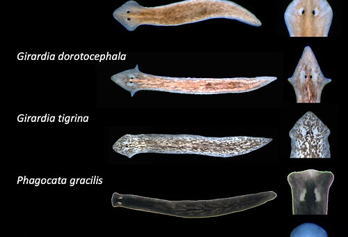
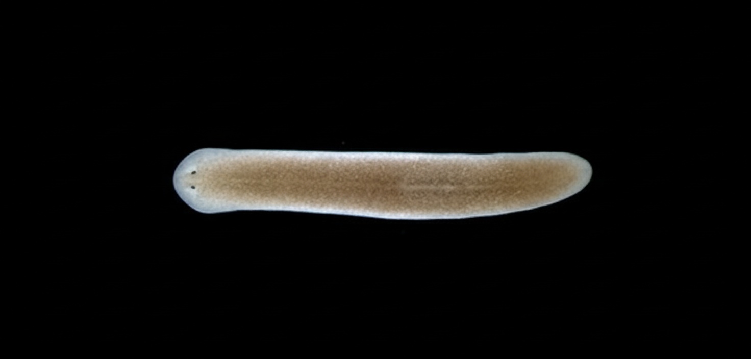
Above: A worm whose cell-to-cell (gap junction) communication was blocked during regeneration, so each of its four wounds grew a head.
What prevents us from growing six eyes, or nine, instead of just two? Why do millions of regenerating planarians look the same, even though they are growing new tissues? The answer: cell signaling! We are interested in how regenerating tissues know what shape to form, which will provide strategies to help regenerative medicine replace lost organs or limbs.
Our Research:
Shape is essential…from proteins to organisms. Shape changes and tissue remodeling drive development, disease and even aging. But despite the importance of shape, we still know very little about how shape is established, maintained, and (following injury) restored. Although recent advances have enabled us to determine developmental pathways and cell fate mechanisms, this still does not explain how changes in individual cells lead to an animal's gross anatomy. So the main questions remain. Why do babies all have that stereotypical “human” shape (and never look like frogs or fish or birds)? How does that human embryo know what “human” should look like, anyway? Our research uses planaria as a model of regenerative shape. Planarians have great plasticity (the ability of tissues to be reshaped) and yet are normally resistant to malformations during regeneration. However, by disrupting cell signaling in the lab, we can regenerate new worms that are wildly misshapen.

Above: Control (bottom center) and H,K-ATPase inhibited (remaining) regenerates illustrate the changes in head shape when membrane voltage is altered.
Summary
How does an amputated fragment of tissue know what shape to become? We exploit the natural diversity in body shape across planarian species to identify molecular and biophysical mechanisms that determine and re-establish animal body morphology during regeneration.
Methods
Comparative transcriptomics across planarian species combined with targeted gene manipulation, bioelectric measurements, and quantitative morphometric analysis allow us to identify shape-determining pathways.
Significance
Revealing how body shape is encoded and restored during regeneration could unlock strategies for rebuilding complex tissues with correct form — a key challenge in regenerative medicine.
Related Publications
Beane, W.S., Morokuma, J., Adams, D.S., Levin, M. (2011). A Chemical Genetics Approach Reveals H,K-ATPase-Mediated Membrane Voltage Is Required for Planarian Head Regeneration. Chemistry & Biology 18(1):77–89.
Beane, W.S., Morokuma, J., Lemire, J.M., Levin, M. (2013). Bioelectric signaling regulates size in zebrafish fins. PLoS Genetics 9(1):e1003236.