The Beane Lab

Stem Cells, Regeneration & Morphogenesis
in the age of Quantum Biology

Beane Lab planarian worm
Wound Epithelium

Wound Epithelium

F-actin (green) and nuclear (blue) staining reveals the morphology of the wound edge.

1/3

Current Research

Stem cells, regeneration, and the physics of life

ROS fluorescent staining in planarian blastema
ROS Signaling

Project 1: ROS Signaling and the Initiation of Regeneration

Summary

Reactive oxygen species (ROS) are short-lived molecules produced after wounding that act as critical second messengers, initiating the cascade of events required for stem cell activation and new tissue growth.

Methods

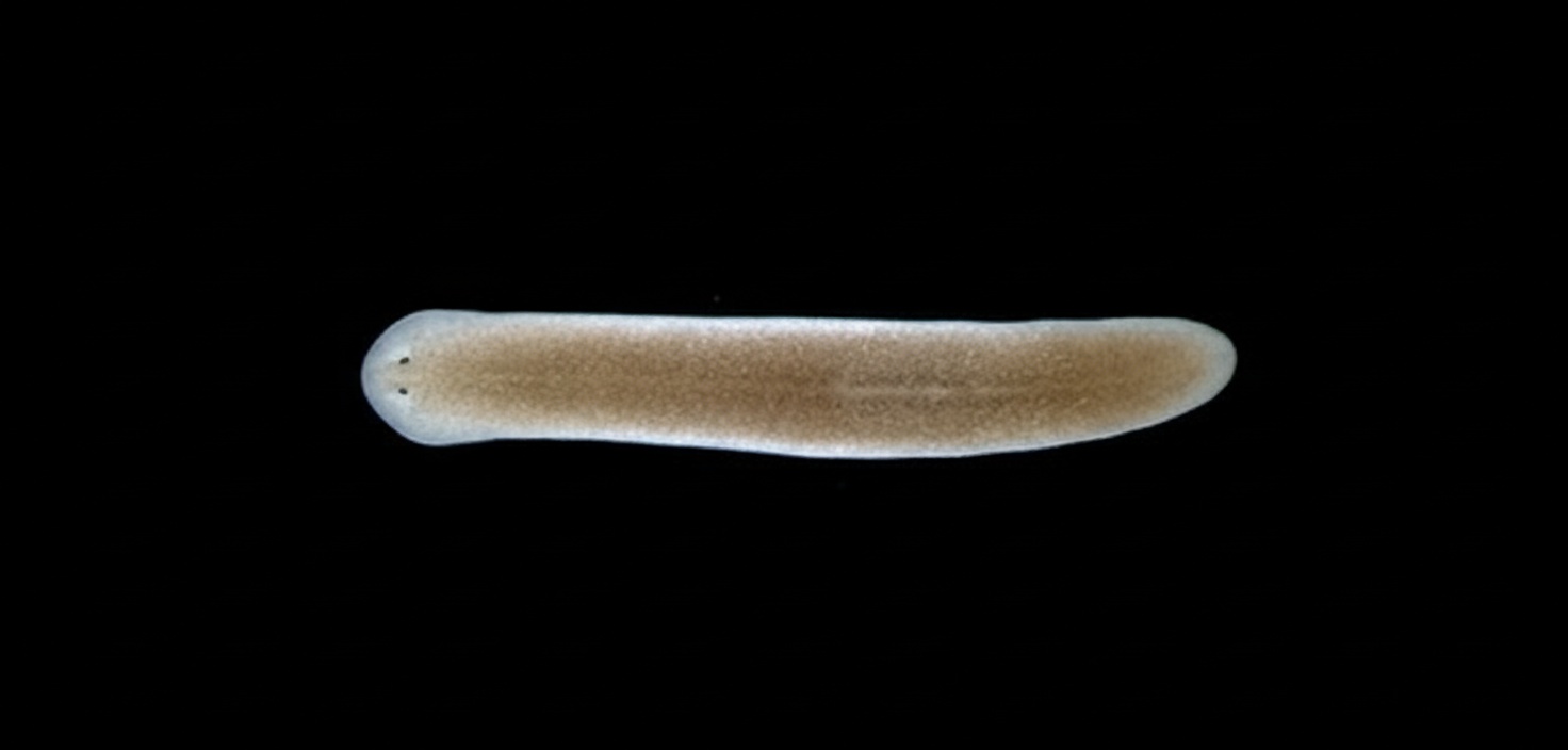
We use fluorescent ROS reporters, live imaging, pharmacological inhibitors, and RNAi knockdown in planarians to dissect the temporal and spatial regulation of ROS during wound healing and blastema formation.

Significance

Understanding how ROS coordinates stem cell proliferation could inform the development of therapies for non-healing wounds and cancer, where ROS signaling is often dysregulated.

Quantum biology magnetic field visualization
Quantum Biology

Project 2: Using Quantum Phenomena to Control Stem Cells

Summary

Radical pair reactions — a quantum mechanical process — may be exploited by biological systems. We investigate whether weak magnetic fields can alter superoxide radical pair recombination rates, thereby modulating ROS levels and, consequently, stem cell behavior.

Methods

We apply precisely controlled weak magnetic fields (0–10 mT) to regenerating planarians and measure effects on superoxide production, stem cell proliferation, and regeneration outcomes through behavioral assays and quantitative imaging.

Significance

If quantum effects on radical pairs can be used to non-invasively control stem cell activity, this opens an entirely new paradigm for regenerative medicine and cancer therapy — control without chemicals.

Synapsin labeling of planarian nervous system
Neural Growth

Project 3: Neural Regeneration and Stopping New Growth

Summary

While much attention has been paid to how neural growth is initiated after injury, our lab asks the opposite question: how does the regenerating nervous system know when to stop growing? We study the planar cell polarity (PCP) pathway’s role in terminating neurogenesis.

Methods

Using RNAi to knock down PCP pathway components in planarians, combined with immunofluorescence for synapsin and other neural markers, we map the spatial and temporal regulation of neural growth termination.

Significance

Aberrant neural growth termination is implicated in neuropathic pain and neural tumors. Identifying stop signals could provide targets for conditions involving runaway neural growth.

Multiple planarian species showing body shape diversity
Morphogenesis

Project 4: Planarians as a Model for Regenerative Shape

Summary

How does an amputated fragment of tissue ‘know’ what shape to become? We exploit the natural diversity in body shape across planarian species to identify the molecular and biophysical mechanisms that determine and re-establish animal body morphology during regeneration.

Methods

Comparative transcriptomics across planarian species combined with targeted gene manipulation, bioelectric measurements, and quantitative morphometric analysis allow us to identify shape-determining pathways.

Significance

Revealing how body shape is encoded and restored during regeneration could unlock strategies for rebuilding complex tissues with correct form — a key challenge in regenerative medicine.